Inflammatory Markers: Ordering and Interpretation


A patient returns with several weeks of fatigue and joint aches, and an earlier visit has already produced a mildly elevated C-reactive protein (CRP). The result then needs interpretation, because the number alone rarely explains the cause or source of inflammation. Inflammatory markers reach this position often, because they are inexpensive and familiar, and that accessibility lets a nonspecific result drive both missed diagnoses and overdiagnosis.
Clinicians order inflammatory markers for triage support, diagnostic framing, treatment monitoring, and risk stratification across infectious, autoimmune, malignant, thrombotic, and inflammatory conditions. The markers in common use, including CRP, high-sensitivity CRP (hs-CRP), the erythrocyte sedimentation rate (ESR), ferritin, procalcitonin, fibrinogen, D-dimer, and several complete blood count (CBC)-derived measures, differ in what they reflect, how fast they move, and what confounds them. This article sets out a framework for ordering and interpreting them, covering CRP versus ESR, marker selection, confounders, monitoring, and when a result should not slow down care.
Ready to start delivering better patient care?
Join 125,000 healthcare providers who rely on Fullscript to dispense top-quality supplements and labs to their patients.
What inflammatory markers measure and what they cannot diagnose
Inflammatory markers can signal an inflammatory response is present and give a rough sense of its magnitude. They do not name its cause, locate its source, or establish a diagnosis on their own.
Inflammation as a biological signal rather than a diagnosis
Many commonly used inflammatory markers are acute-phase reactants, plasma proteins whose concentration shifts when inflammation is present. The liver increases their production under cytokine stimulation, particularly interleukin-6 (IL-6), in response to infection, tissue injury, autoimmunity, malignancy, trauma, or metabolic stress. (4) A rising marker shows that something inflammatory is underway without indicating where, because systemic and localized processes can produce similar values.
Broad inflammatory markers differ from disease-specific tests such as autoantibodies, cultures, serology, imaging, and biopsy. A nonspecific marker can raise or lower suspicion, while a specific test assigns the cause. Against a low pretest probability an abnormal result is more likely a false positive and can set off low-yield imaging and repeat testing, while against red flags a normal result can falsely reassure, so the decision to order, observe, test further, or escalate comes before the result. Pretest probability is the likelihood of a condition before the marker result is known. A false positive is an abnormal result in a patient without the condition being considered, a false negative is a normal result despite the condition being present, and diagnostic specificity describes how well a test avoids positive results in patients without the condition.
Appropriate clinical roles for inflammatory markers
Inflammatory markers are useful in a few defined roles. The first is screening support when the clinical picture already suggests an inflammatory, infectious, rheumatologic, or malignant process, where a symptom pattern justifies a marker and routine ordering in a low-risk patient mostly surfaces incidental findings. The second is monitoring known disease activity, in rheumatologic conditions, inflammatory bowel disease (IBD), a treated infection, or a postoperative course, where the trend across serial values is more informative than any single result. (11) The third is risk stratification and triage when serious infection or a systemic, thromboinflammatory, or hyperinflammatory syndrome is under consideration, where an unstable patient may need escalation independent of any marker value.
Common misuses and overinterpretation
Ordering a broad panel without a clinical question is a frequent misuse. Drawing CRP, ESR, ferritin, D-dimer, fibrinogen, procalcitonin, and selected CBC-derived measures together by reflex raises the chance of an incidental abnormality and downstream testing, and an isolated mild elevation without concerning history or examination findings does not justify an extensive workup. (13) Treating an abnormal result as a diagnosis is a second misuse, since "high inflammation" describes a state, not a cause, a location, or a severity, and reading a nonspecific elevation as autoimmune disease leads to false-positive cascades and anchoring on the first result. Treating a normal result as exclusionary is a third, because early, localized, intermittent, or immunomodulated disease can run with a limited marker response and serious disease can show little initial change, so red flags take priority over a reassuring marker.
How to interpret inflammatory markers: a clinician framework
Interpretation of inflammatory markers follows four questions:
- What is the clinical question? The reason the marker was ordered, whether diagnosis, triage, monitoring, or risk stratification, sets what the result can do, and a test without a defined use cannot change management.
- What is the pretest probability? Symptoms, examination, and the syndrome pattern decide whether an abnormal value is more likely true or false, so a low-probability result risks a false positive and one read against red flags risks false reassurance.
- What do timing, trend, and confounders show? Acute and chronic presentations behave differently, prior values establish a baseline, and recent infection, trauma, surgery, vaccination, comorbidities, and medication all change what to expect.
- Does the result change care? A useful marker result should affect targeted testing, imaging, monitoring, referral, escalation, or observation, while one that supports neither diagnosis nor reassurance changes nothing.
Marker-by-marker clinical utility
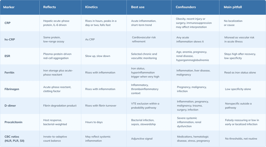
The common markers differ in what they reflect, how fast they move, and what confounds them.
CRP and hs-CRP
C-reactive protein is an acute-phase reactant produced by the liver under interleukin-6 stimulation, able to climb steeply during infection or tissue injury. (4) CRP begins to rise within hours and peaks within about a day or two, and its short half-life ties the level to current production, so a trend is more informative than a single result when the baseline was elevated. (11) It does not localize inflammation or distinguish infection from noninfectious causes such as trauma, surgery, or autoimmune disease, which act through the same acute-phase response. (4)
High-sensitivity CRP measures the same protein with a low-range assay used in cardiovascular risk assessment, where in adults at intermediate risk hs-CRP is one of the risk-enhancing factors that can inform a statin decision,with more recent guidance recommending broader hs-CRP screening in both primary and secondary prevention. (1) Drawn during an acute infection or flare it reflects that process and carries little meaning for baseline vascular risk. Outside acute infection, obesity is associated with higher CRP, since adipocytes are among the cells that produce the protein, and recent infection, trauma, surgery, or vaccination can keep it elevated while immunosuppression may blunt the rise, so a single value is best read against timing, comorbidities, and prior results. (12)
ESR
The erythrocyte sedimentation rate is an indirect marker, the distance red cells fall in a tube over an hour. Plasma proteins, fibrinogen in particular, reduce the surface charge on red cells and promote aggregation, raising the rate, and ESR moves more slowly than CRP and stays elevated longer, suiting selected chronic and vasculitic contexts while limiting its specificity. (4) Age, pregnancy, anemia, renal disease, and hypergammaglobulinemia raise the ESR independent of inflammation, and a delay before the sample is run can alter it. (13) It can also stay elevated after clinical improvement, and a very high ESR tends to accompany identifiable serious disease such as infection, malignancy, or temporal arteritis, while a mild elevation without concerning findings does not justify an extensive search or replace disease-specific testing when rheumatologic disease is suspected. (13)
Ferritin, fibrinogen, and D-dimer
Ferritin is both an iron-storage protein and a positive acute-phase reactant, so its level reflects iron status and inflammation at once and is interpreted with iron studies when anemia or overload is in question. (4) A markedly elevated ferritin with systemic symptoms widens the differential to infection, liver disease, malignancy, and hyperinflammatory syndromes, so an isolated value without context is hard to interpret. (8) Fibrinogen is an acute-phase reactant and clotting factor that rises in inflammatory and thromboinflammatory states and in pregnancy, malignancy, and infection, with limited specificity alone. (4) D-dimer, a fibrin degradation product, rises in venous thromboembolism (VTE) but also in inflammation, pregnancy, malignancy, trauma, infection, and postoperative states, so it is informative only within a validated probability pathway, where a value below a fixed, age-adjusted, or probability-adjusted threshold helps exclude VTE at a nonhigh probability. (3)
Procalcitonin
Procalcitonin supports the assessment of selected bacterial infection and sepsis as an adjunct to clinical judgment, cultures, imaging, and source evaluation, and is most useful within an antibiotic-stewardship framework, with sensitivity and specificity imperfect enough that interpretation in clinical context is required. (10) Serial values are read against the clinical trajectory, and antibiotic decisions should not rest on procalcitonin alone, with institutional protocols governing use. (10) It can read falsely high in severe systemic inflammation, low or falsely reassuring in early or localized infection, and is further limited in nonbacterial inflammation, immunomodulated states, and renal dysfunction.
CBC-derived markers and composite indices
Components of the complete blood count carry inflammatory clues, since leukocytosis, neutrophilia, lymphopenia, eosinophilia, thrombocytosis, and the anemia of chronic inflammation can each accompany inflammation, all read alongside the smear, differential, and syndrome. The neutrophil-to-lymphocyte ratio (NLR), platelet-to-lymphocyte ratio (PLR), and systemic immune-inflammation index (SII) combine routine counts into measures of systemic inflammation, and they are inexpensive and accessible, but definitive thresholds are not established and their role remains adjunctive rather than routine. (5) Medication effects, hematologic disease, an acute stress response, and pregnancy all shift these counts, so interpretation stays syndrome-specific.
CRP vs ESR in clinical interpretation
CRP and ESR both rise with inflammation, but they move on different timescales and answer slightly different questions.
Kinetics and use-case differences
CRP rises within hours and falls quickly once inflammation resolves, because its short half-life ties the level to current production. (11) ESR changes more slowly and lags behind clinical change, and baseline and prior values matter for both. (13) In acute infection and tissue injury CRP tracks the process closely, in some chronic inflammatory disease and in vasculitis and polymyalgia rheumatica (PMR) ESR retains a role, and in suspected giant cell arteritis (GCA) both tests together can improve diagnostic interpretation compared with either alone. (6) For monitoring, a CRP trend signals near-term change while ESR can lag, and disease-specific activity measures are the preferred anchors where they exist.
Discordant CRP and ESR results
A high CRP with a normal ESR can suggest an early or acute process the slower ESR has not yet reflected, read against timing and trajectory. A high ESR with a normal CRP points toward age, anemia, pregnancy, renal disease, or a plasma-protein abnormality, a chronic inflammatory or malignant process when the picture supports it, or an ESR still elevated after inflammation has resolved, and it suits targeted follow-up more than broad testing. (13) A normal CRP alongside symptoms of inflammation occurs in localized, early, intermittent, or medication-modified disease, and red-flag symptoms warrant evaluation despite normal markers.
Practical CRP vs ESR ordering strategy
CRP alone may suffice for acute inflammatory assessment, short-interval monitoring, and treatment-response tracking when it tracks the disease state, and a duplicate ESR adds little when it will not change management. ESR adds value in selected vasculitic and rheumatologic presentations, chronic inflammatory patterns, a plasma-protein or hematologic context, and where prior ESR trends guide specialty care, and in suspected GCA both tests are commonly drawn. (6) Neither is the right next step when suspicion is high for a specific infection (cultures or imaging), autoimmune disease (serology), thromboembolism (a validated probability pathway), or malignancy (a syndrome-directed evaluation).
How to order and interpret inflammatory markers by clinical question
The same marker means different things depending on the question that prompted it. The sections that follow work through the common clinical questions.
Fever or suspected infection
Severity, hemodynamics, localizing symptoms, and immune status drive whether cultures, imaging, source control, or empiric therapy come first. CRP supports the inflammatory burden, a CBC with differential shows the host-response pattern, procalcitonin is added only when clinically and institutionally appropriate, and D-dimer is avoided unless a thromboembolic probability pathway is in play. (10) The safety boundary is firm: sepsis evaluation and management are not delayed for marker confirmation, and for septic shock or a high likelihood of sepsis antimicrobials are given immediately, while when sepsis is possible without shock a rapid assessment is performed and antimicrobials are administered within 3 hours if the concern for infection persists. (9) Normal early markers do not exclude serious infection in an unstable patient.
Chronic joint pain or suspected rheumatologic disease
An inflammatory pain pattern, prolonged morning stiffness, synovitis with functional limitation, and systemic features such as fever, weight loss, or rash drive testing. CRP and ESR support the inflammatory burden, disease-specific serology is added when indicated, and imaging follows when joint disease is suspected, but a marker pattern does not assign an autoimmune diagnosis on its own. Suspected inflammatory arthritis or vasculitis, constitutional symptoms with abnormal markers, or organ-threatening progression warrant specialist involvement.
Monitoring known inflammatory disease
In autoimmune and rheumatologic conditions, marker trends are read against an individual baseline, symptoms, examination, and validated activity measures, since markers and clinical activity can diverge. In inflammatory bowel disease, CRP and fecal calprotectin are used as supportive treat-to-target markers, with calprotectin generally more sensitive than CRP or ESR for intestinal mucosal activity, and trends integrate with endoscopy and symptoms rather than standing alone, so marker-only escalation or de-escalation is avoided. (7) In infection follow-up, serial markers are drawn only when they will change management and do not replace reassessment.
Elevated ferritin with systemic symptoms
A marked ferritin elevation with systemic symptoms spans acute or chronic inflammation, infection, liver disease, malignancy, iron overload, and hyperinflammatory syndromes such as hemophagocytic lymphohistiocytosis (HLH) and macrophage activation syndrome (MAS). (8) Useful co-testing includes a CBC with differential, liver enzymes, and iron studies, adding triglycerides and fibrinogen when hyperinflammation is suspected. Systemic illness with cytopenias, organ dysfunction, coagulopathy, or persistent fever alongside very high ferritin should prompt urgent hematology or inpatient evaluation, since adult HLH carries high mortality and benefits from early referral, and ferritin alone is not a basis for outpatient reassurance here. (2)
High CRP in an otherwise asymptomatic patient
Recent infection or vaccination, trauma, surgery, exertion, obesity, smoking, or metabolic disease can each raise CRP, so a medication review and a repeat test when justified come before further workup. Broad autoimmune panels without symptoms and imaging cascades without localizing findings tend to generate false positives, and an isolated mild elevation without concerning findings does not justify an extensive workup. (13) Persistent or rising values, new or constitutional symptoms, abnormal basic labs, or risk factors for cancer, infection, or inflammatory disease move the evaluation forward.
Confounders, false positives, and false reassurance
Many values that look abnormal reflect a confounder rather than the disease under consideration, and many normal values sit alongside real disease.
Patient-level confounders
Age and pregnancy shift several markers, and baseline variability differs between people. Anemia, polycythemia, hypergammaglobulinemia, chronic kidney disease (CKD), and altered plasma-protein states change the ESR and other markers independent of inflammation. (13) Obesity, smoking, metabolic comorbidities, and chronic cardiovascular disease can raise CRP or hs-CRP baseline levels, while recent strenuous exertion can transiently affect inflammatory markers.
Clinical event confounders
Surgery, trauma, burns, and procedure-related inflammation raise markers along an expected recovery trajectory, so timing relative to the event guides interpretation. A recent infection leaves a convalescent lag, and inflammation can persist after clinical improvement, blurring the line between relapse and slow normalization. Cancer and chronic renal, hepatic, or hematologic comorbidity can also keep markers elevated, so not every elevation in a patient with a known diagnosis belongs to that diagnosis.
Medication and treatment effects
Glucocorticoids, biologic agents, and Janus kinase (JAK) inhibitors and other targeted therapies can blunt the inflammatory response, so a normal or modest marker in an immunosuppressed patient does not exclude serious infection. Nonsteroidal anti-inflammatory drugs (NSAIDs) may ease symptoms without resolving the process, and antibiotics started before testing can alter values, so results are read against timing. D-dimer is interpreted with care in anticoagulated patients, and hs-CRP for cardiovascular risk belongs to a prevention context separate from acute inflammatory evaluation, so the two uses are not conflated. (1)
Red-flag contexts where markers must not delay care
In several settings the clinical picture, not the marker, drives the decision, and waiting for a result can cause harm.
Suspected sepsis or serious infection
Hypotension, tachypnea or hypoxia, altered mental status, persistent fever, and organ dysfunction call for action regardless of a marker. CRP and procalcitonin offer adjunctive risk and trend support, and cultures, imaging, and source evaluation follow as indicated, but empiric management proceeds on clinical severity, and antimicrobials for septic shock or a high likelihood of sepsis are not held for a marker result. (9)
Suspected giant cell arteritis or polymyalgia rheumatica
A new headache in an older adult, jaw claudication, visual symptoms, scalp tenderness, and proximal girdle pain and stiffness are the high-risk features. CRP and ESR support the assessment and ordering both reduce the chance of missing GCA compared with relying on either alone. However, a small proportion of biopsy-proven GCA have normal inflammatory markers at diagnosis, so normal results do not eliminate concern when suspicion is high. (6) Visual or ischemic symptoms warrant urgent specialist evaluation, and the diagnosis rests on a disease-specific pathway.
Suspected HLH or other hyperinflammatory syndromes
Persistent fever, cytopenias, hepatosplenomegaly, organ dysfunction, coagulopathy, and a very high ferritin form the pattern, with evaluation for an infectious, malignant, or rheumatologic trigger. Compatible features warrant urgent specialist involvement, since adult HLH carries high mortality and benefits from early referral, and an isolated ferritin threshold without the full clinical context does not establish the diagnosis. (2)
Severe inflammatory bowel disease flare or complication
Severe abdominal pain, fever, tachycardia, bloody diarrhea, dehydration, peritoneal signs, and anemia are the red flags. CRP and other markers support a severity assessment and integrate with stool testing, imaging, endoscopy, and complication assessment, and a normal or modest marker does not justify outpatient reassurance when clinical severity is high. (7)
Interpreting patterns without overtesting
Timing, prior values, and the pattern across markers often add more than additional nonspecific tests.
Acute versus chronic inflammation patterns
A rapid symptom onset with a rising CRP can support an acute inflammatory process such as infection, trauma, surgery, or a flare, and time-sensitive diagnoses take priority. (11) Persistent elevation can accompany chronic inflammatory disease, malignancy, renal disease, or metabolic inflammation, and targeted evaluation suits it better than repeated broad panels. Episodic symptoms may be better assessed with testing during a flare, with fever syndromes, autoinflammatory disease, or relapsing infection in the differential.
Choosing targeted testing over broad panels
A localizing history, the organ-system pattern, exposure history, and a medication and immune-status review select the disease-specific pathway: cultures or molecular testing for infection, autoantibodies or complement studies for rheumatologic disease, imaging or endoscopy for localization, and biopsy for a tissue diagnosis. A marker is repeated only when timing and trajectory matter, and trending stops once the result no longer changes care.
Common misinterpretations in clinician use
The belief that markers diagnose autoimmune disease overstates the test, since markers adjust probability but do not define disease, which requires the syndrome pattern and disease-specific testing. The belief that a normal CRP rules out serious disease is unsafe, because early, localized, immunomodulated, or intermittent disease can show a limited marker response while the protein takes time to rise, and red flags override marker reassurance. (11) The belief that a high CRP always means infection ignores its biology, since noninfectious inflammation, trauma, malignancy, and autoimmune disease all raise CRP, and infection likelihood depends on the syndrome, exposure, immune status, and cultures. (4) The belief that more markers improve accuracy backfires, because broad panels raise the chance of an incidental abnormality while targeted testing is more likely to answer the clinical question. (13)
Clinician communication, documentation, and follow-up
How a marker result is explained and recorded shapes what happens next as much as the value does.
Clinician communication about "high inflammation"
An inflammatory marker reports a biologic response, not a diagnosis, read with symptoms, examination, timing, and comorbidities, with the next step depending on whether it changes management. Naming "autoimmune disease," "infection," or "cancer" on a marker alone overstates the test, so communicating uncertainty without dismissing symptoms keeps the conversation accurate. When markers are normal but symptoms persist, the normal value does not invalidate the symptoms, and follow-up depends on severity, persistence, red flags, and clear safety-net instructions.
Documentation standards for marker interpretation
Useful documentation records the clinical question: the reason for ordering, the differential, and whether the marker supports diagnosis, triage, or monitoring. For an abnormal result, it captures the acute or chronic context, relevant confounders, prior comparison values, and the action taken or reason for observation. It also records the escalation triggers, including the symptoms that should prompt urgent reassessment and the disease-specific testing or referral plan.
Frequently asked questions
What do inflammatory markers show in clinical practice?
Inflammatory markers can signal an inflammatory response is present and give a rough sense of its magnitude, but they do not identify the cause, locate the source, or establish a diagnosis without history, examination, and disease-specific testing.
How should clinicians interpret inflammatory marker results?
A result is read against the clinical question, the pretest probability, the timing and trend, and the confounders that apply, and what counts is whether it changes the differential, the urgency, or the plan.
When should clinicians order CRP and ESR together?
Both are commonly drawn when giant cell arteritis or polymyalgia rheumatica is suspected, since ordering both reduces the chance of missing GCA compared with relying on either alone, while in many acute settings CRP alone answers the question.
What is the practical difference between CRP and ESR?
CRP rises within hours and falls quickly once inflammation resolves, so it tracks short-term change, while ESR moves more slowly, can stay elevated after improvement, and is influenced by age, anemia, pregnancy, and plasma proteins.
How should an elevated CRP be interpreted when the patient is otherwise asymptomatic?
Recent infection, vaccination, injury, surgery, obesity, and smoking can each contribute to an elevated CRP, so context and a repeat test come before further workup, and an isolated mild elevation without concerning findings does not justify an extensive evaluation.
What does high ESR with normal CRP suggest?
This pattern can often reflect age, anemia, pregnancy, renal disease, a plasma-protein abnormality, or inflammation that has resolved rather than active inflammation, and it suits targeted follow-up more than broad testing.
Can normal inflammatory markers exclude autoimmune disease?
No. Early, localized, intermittent, or medication-modified disease can run with normal markers, and an autoimmune diagnosis depends on the syndrome pattern and disease-specific testing.
Which inflammatory markers are most useful for suspected infection?
CRP supports the inflammatory burden and its trend and a CBC with differential shows the host-response pattern, while procalcitonin can help within an antibiotic-stewardship framework despite imperfect sensitivity and specificity.
When is procalcitonin appropriate, and when can it mislead?
Procalcitonin supports selected bacterial infection and sepsis assessment as an adjunct to clinical judgment, cultures, and imaging, but it can read falsely high in severe systemic inflammation and falsely low in early or localized infection, so antibiotic decisions should not rest on it alone.
How should ferritin be interpreted when systemic symptoms are present?
Ferritin is both an iron-storage protein and an acute-phase reactant, so a markedly elevated value with systemic symptoms widens the differential to infection, liver disease, malignancy, and hyperinflammatory syndromes, and persistent fever, cytopenias, or organ dysfunction alongside very high ferritin warrants urgent evaluation rather than reassurance.
How should inflammatory markers be used to monitor treatment response?
Marker trends are read against an individual baseline and alongside symptoms, examination, and validated activity measures, and in inflammatory bowel disease CRP and fecal calprotectin serve as supportive targets that integrate with endoscopy and do not stand alone.
Which confounders most commonly cause false-positive inflammatory marker results?
Age, pregnancy, anemia, renal disease, obesity, and recent infection, trauma, or surgery commonly raise markers independent of the disease under consideration, with plasma-protein abnormalities raising the ESR in particular.
When should inflammatory markers prompt urgent escalation rather than outpatient follow-up?
When red flags are present, such as instability suggesting sepsis, visual or ischemic symptoms suggesting giant cell arteritis, persistent fever with cytopenias and very high ferritin, or a severe inflammatory bowel disease flare, the clinical picture drives urgent evaluation regardless of the marker value.
The bottom line
Inflammatory markers are most useful when ordered to answer a defined clinical question and read against pretest probability, the syndrome pattern, comorbidities, medications, and timing. CRP, hs-CRP, ESR, ferritin, procalcitonin, fibrinogen, D-dimer, and CBC-derived measures each carry their own interpretation logic, and treating them as interchangeable readouts of inflammation loses that detail. The safest frame for a result is not normal versus abnormal but whether it changes the differential, the urgency, the monitoring plan, or the need for disease-specific evaluation. Ordering built around the clinical syndrome, the expected effect on management, confounders, red-flag screening, and a documented follow-up threshold keeps inflammatory markers in the supporting role they fill best.
Ready to start delivering better patient care?
Join 125,000 healthcare providers who rely on Fullscript to dispense top-quality supplements and labs to their patients.